Our Clinic
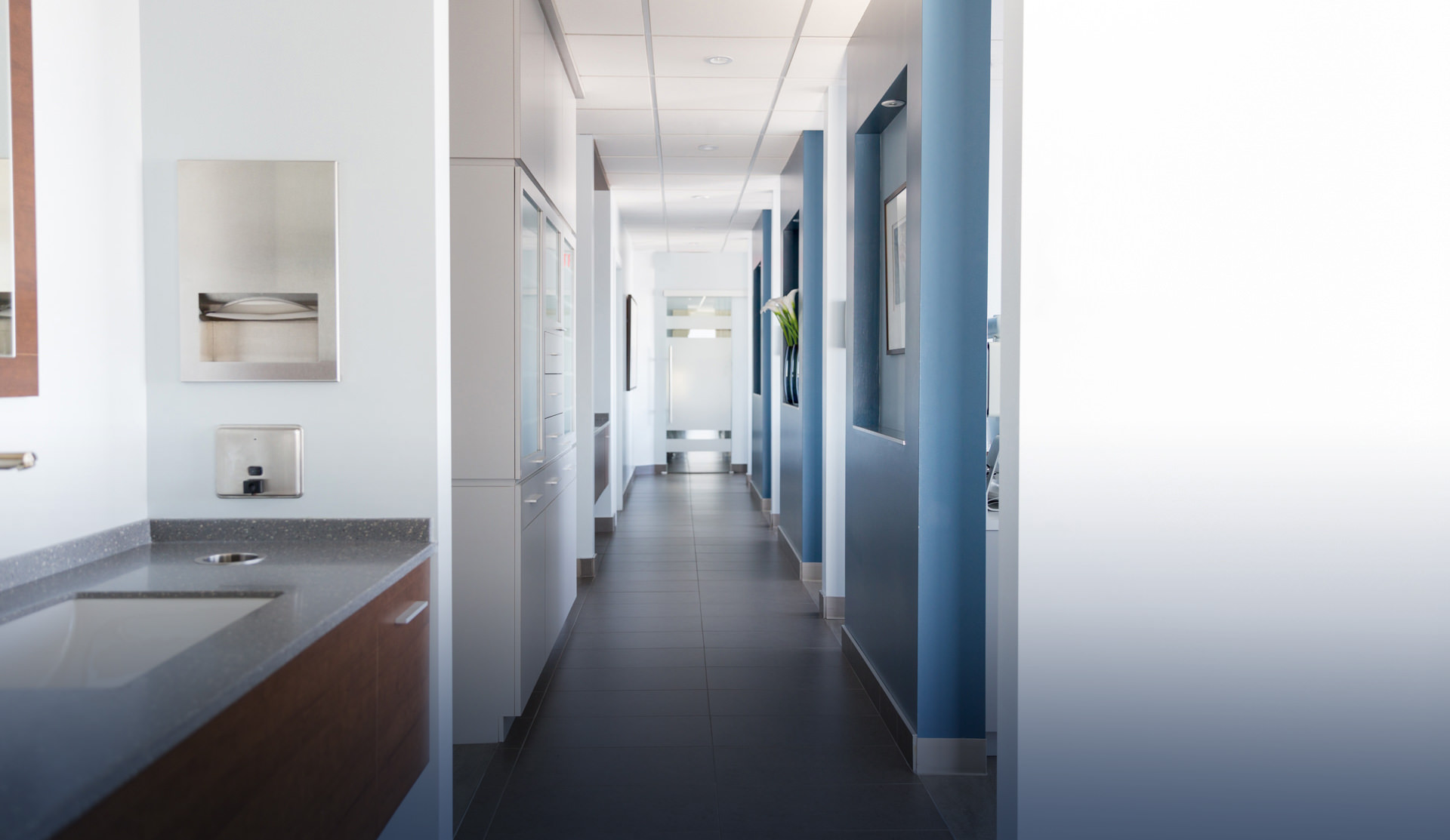
We are dedicated to excellence in the art and science of endodontics. Here are some cutting-edge technologies that are essential to us in providing the best of modern, minimally invasive endodontics.
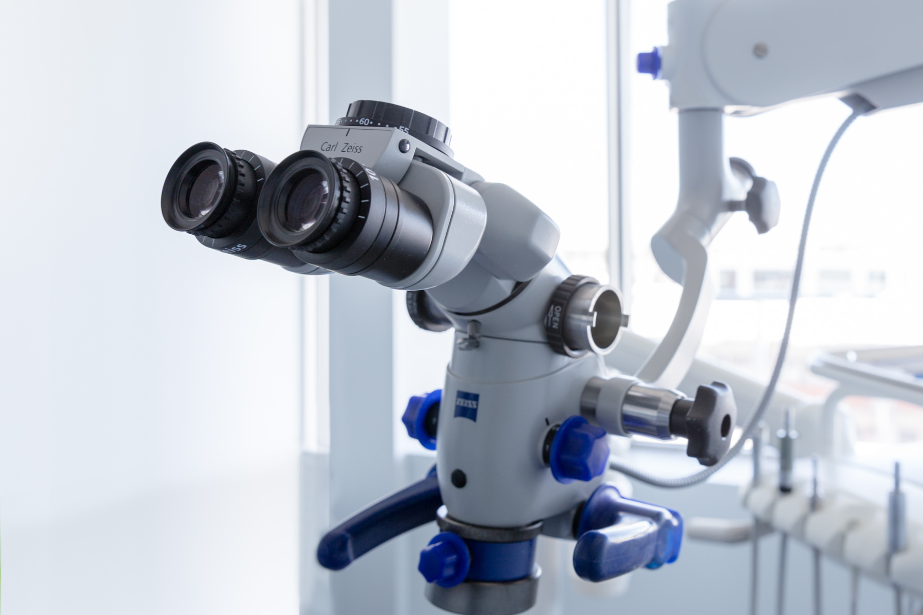
Clinical Microscope
The clinical microscope is an essential and irreplaceable part of the basic equipment of the endodontist. This particular equipment offers a magnified image of X5 to X25 times of the actual size of the teeth which allows to:
- Find canals
- Reduce the size of the access cavity, the whole made inside the tooth so as to endodontically treat it.
- Confirm diagnosis (fracture, perforation or resorption)
- Be more precise when doing a surgical approach for apectomies
The recent literature reports higher success rates when using a clinical microscope and therefore insures endodontic treatments which are reproductible and reliable.
Each of our 6 operating rooms are equipped with a clinical microscope Zeiss, a renowned brand in the world of optics.

Endodontic Laser
At our endodontic clinic we use the Fotona LightWalker® II for SWEEPS® (Shock Wave Enhanced Emission Photoacoustic Streaming)—a laser method that energizes the irrigation solution inside the tooth. Brief laser pulses create micro-bubbles; their gentle collapse generates fluid motion that carries the irrigant into fins, isthmuses, and lateral canals that files can’t reach.
How we use the laser in root canal care:
- Primary treatment: enhances cleaning of complex anatomy, especially in the apical third.
- Retreatment: helps disrupt biofilm and remove debris around old materials.
- Conservative shaping: supports thorough disinfection with less aggressive instrumentation.
- Workflow: efficient rinsing between steps and a clean field for sealing.
What this means for patients: more thorough chemomechanical cleaning in a focused appointment, with techniques designed to support comfortable recovery and long-term tooth preservation.
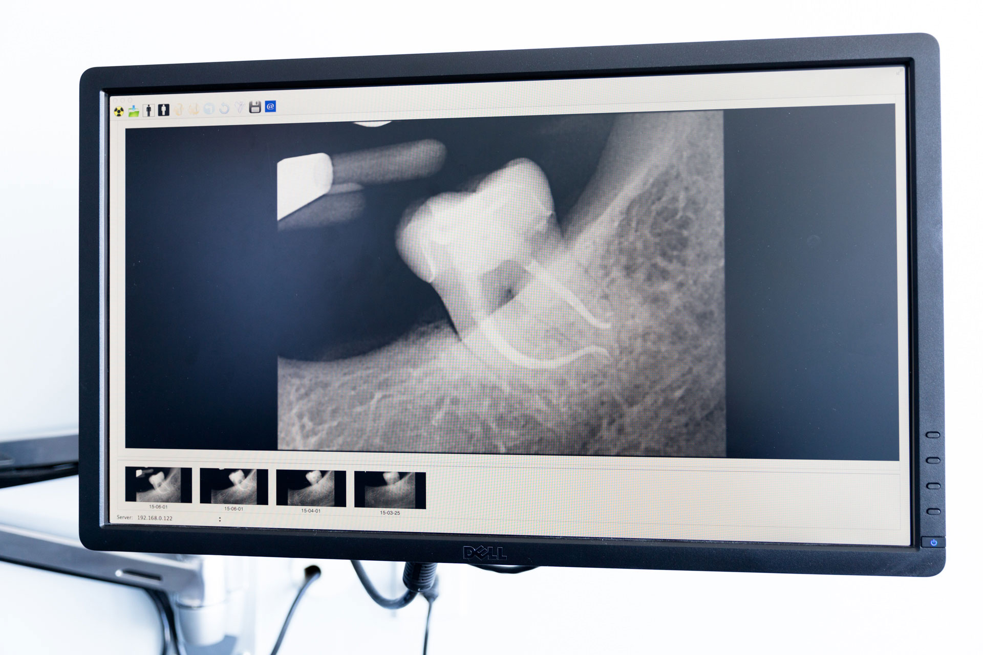
Digital Radiography
Each of our 6 operating rooms are equipped with digital radiography.
Digital radiography allows:
- To significantly reduce the amount of exposure ti x-rays.
- To better visualize the image on a wider screen and therefore facilitate the information given during your clinical exam and during the treatments.
- To facilitate the transmission and the sharing of the images with your dentist.
- The usage of an ecologically friendly technology which is respectful of the environment, since this method diminishes the usage of chemical products.
.jpg)
CBCT
Dental anatomie varies greatly from one patient to the next. The endodontist prior to treating a tooth attempts to understand the specific anatomy of each tooth to be treated, as well as its roots and the bone surrounding it. Conventional radiographs offer a limited 2-dimension perspective on the area examined. With modern endodontics, comes also the introduction of the Cone-beam Computed Tomography, often called CBCT. This technology offers a 3D image of the examined tooth. This 3D scan give us a lot of information on the tooth and surrounding tissus. This moderately recent technology allows to reliably plan the endodontic treatment. While it is necessary in all cases, it can be useful in cases of pretreatment, apical surgery, complex diagnostic examination or dental trauma cases.
In addition to our digital radiography, we have a CBCT machine in our office since 2013. Our unit take a 3D radiographic image of a limited region of 3-4 teeth. This allows an increased precision and limits the exposure to x-rays.


Office
Town of Mount Royal, QC H3P 3H5